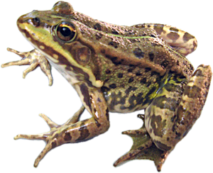
Bondareva et al. (2012) Cytogenetic characteristics of erythrocytes of green frogs
Bondareva A. A., Bibik Yu. S., Samilo S. M., Shabanov D. A. Cytogenetic characteristics of erythrocytes of green frogs from the Siversky Donets diversity centre of the Pelophylax esculentus complex // Bulletin of V. N. Karazin Kharkiv National University. Series: Biology. – 2012. – Issue 15 (No. 10...
Cytogenetic features of erythrocytes of green frogs from the Siversky Donets center of the Pelophylax esculentus complex diversity A.A.Bondarieva, Yu.S.Bibik, S.M.Samilo, D.A.Shabanov The influence of triploidy on erythrocyte morphological parameters of Pelophylax esculentus, hemiclonal hybrids of green frogs, is considered. It is shown that triploidy leads to enlarging of erythrocytes while nuclear-cytoplasmic ratio remains constant and relative cell surface decreases. Triploid erythrocytes are characterized by more ellipsoid shape in comparison with diploid cells. Due to erythrocyte size investigated Pelophylax esculentus were divided into three groups: individuals with abnormally small erythrocytes, typical diploids and triploids. The heterochromatin granules quantity in frogs erythrocyte nuclei was estimated. It is supposed that erythrocyte nuclei of individuals with abnormally small cells contain relatively more heterochromatin. Key words: Pelophylax esculentus (= Rana esculenta), erythrocytes, triploids, diploids, heterochromatin. Introduction The green frog hybrid complex (Pelophylax esculentus complex) includes two parental species: the pool frog, Pelophylax lessonae (Camerano, 1882), and the lake frog, Pelophylax ridibundus (Pallas, 1771), as well as their hybrids (named similarly to the species): the edible frog, Pelophylax esculentus (Linnaeus, 1758). Pelophylax esculentus exhibits unusual reproductive strategies, including hemiclonal inheritance – clonal transmission of one of the parental genomes into gametes. In the Siversky Donets center of green frog diversity (Shabanov, Litvinchuk, 2010), both diploid and triploid P. esculentus are widespread. Among them are individuals with two genomes of P. ridibundus and one of P. lessonae, and individuals with two genomes of P. lessonae and one of P. ridibundus. Diploid and triploid P. esculentus live in the same local environments, reproduce in shared breeding groups, have similar sizes, do not significantly differ in lifespan, and have no distinct external or behavioral features (Plotner, 2005; Shabanov, Litvinchuk, 2010; Usova, 2010). This makes them an excellent object for studying the influence of cell ploidy on various cytological parameters. Modern batrachology knows several groups of amphibians that include closely related di-, tri-, and tetraploid species, as observed, for example, in the Bufo viridis complex (Litvinchuk et al., 2011). However, in such groups, individuals with different ploidy belong to different species, differing in their habitats and having undergone more or less independent evolution. The genomes of triploid green frogs have evolved within triploid organisms, while the genomes of triploid green frogs are inherited from diploid parental species. It can be expected that the cytological effects caused by triploidy will be more pronounced in P. esculentus, not being compensated by natural selection. The observed properties of organisms, including their cell characteristics, are the result of a compromise between the needs of optimizing different parameters (Bigon et al., 1989; Rasnitsyn, 2002). In triploid frogs, genomes that are the result of the evolution of diploid organisms are found in triploid nuclei. An interesting research task is to determine how this will affect the nuclear-cytoplasmic ratio, the relative surface area of cells, their number in individual organs and the organism as a whole, and other cell and organism parameters. Diploid and triploid P. esculentus are practically indistinguishable at the biochemical level, as they develop under the control of the same genomes inherited from the parental species. They are also very similar at the organismal level (Plotner, 2005; Usova, 2010). However, they differ sharply at the cellular level, which is a consequence of the different number of parental genomes per cell. This is primarily due to the fact that to maintain the constancy of the nuclear-cytoplasmic ratio in triploids, whose nucleus is 1.5 times larger than in diploids, the cytoplasm content, and thus the cell size, must also increase after the nucleus (Plotner, 2005; Shabanov, Litvinchuk, 2010). A significant difference in erythrocyte size has been recorded for diploid and triploid P. esculentus, and this is used as one of the methods for determining frog ploidy (George, Lennartz, 1980; Polls Pelaz, Graf, 1988; Plotner, 2005). Detailed studies of the influence of frog ploidy on other hematological parameters have not been conducted, whereas for some fish species, it has been shown that triploidy affects various erythrocyte morphological parameters to varying degrees. For example, the major axis of the cell in the studied fish increases to a greater extent than the minor axis (Cal et al., 2005). With increasing cell size, a decrease in their relative surface area is expected (cell volume increases proportionally to the third power of linear dimensions, while surface area increases more slowly, proportionally to the second power). This means that substance transport through the erythrocyte surface in triploid individuals may be impaired. How this circumstance will affect gas exchange is unknown. In our study, we compared erythrocytes of di- and triploid hybrids of green frogs (Pelophylax esculentus). The first level of erythrocyte comparison was the study of their size and morphology, and the second was the comparison of the degree of chromatin condensation. Materials and Methods We studied a sample of frogs collected in the Siversky Donets river basin in the Zmiiv district of the Kharkiv region on September 18, 2010. The sample of 58 individuals, identified by external characteristics (Shabanov et al., 2006) as P. esculentus (body length 6.91±0.62 cm), consisted mainly of males (54 individuals). For each individual, a blood smear was prepared using a standard method and photographed unstained under high magnification (?40) using a USB camera. An object micrometer was photographed under the same magnification. Using the PDF-XChange Viewer program, the major and minor axes of erythrocytes, as well as the major and minor axes of their nuclei, were measured from the smear photographs for 10 cells per smear; these measurements were then converted to micrometers. The cross-sectional area of the cell (visible in the preparation) was calculated using the formula, the surface area of the cell was calculated as the surface area of an ellipsoid of revolution, and the volume using the formula, where a and b are the semi-major and semi-minor axes of the erythrocyte, respectively. Similarly, the cross-sectional area, surface area, and volume of the nucleus were calculated. Based on these data, the nuclear-cytoplasmic ratio was calculated. For 14 individuals from this sample, ploidy, as well as belonging to Pelophylax esculentus, was accurately determined by S. N. Litvinchuk and Yu. M. Rozanov (Institute of Cytology RAS, St. Petersburg) by measuring the amount of DNA per cell using flow DNA cytometry (the method is described, for example, in Litvinchuk et al., 2011). To compare the degree of heterochromatin condensation in erythrocytes, prepared blood smear slides were stained using the Feulgen method (staining with Schiff's reagent after cold hydrolysis with 5N HCl for 20 min) (Roskina, Levinson, 1957). The number of heterochromatin granules was counted under high magnification (?640). A total of 11 blood smears were analyzed (50 cells per smear), including 6 diploid, 3 triploid, and 2 individuals with abnormally small erythrocytes (see below). To determine the degree of heterochromatin decondensation in erythrocyte nuclei, different times of acid hydrolysis were used. For this study, two parts of the same smear of erythrocytes from a specific individual were stained using the Feulgen method with different exposure times in hydrochloric acid (20 min and 60 min). The optical density of nuclei and erythrocyte cytoplasm was determined from photographs of stained smears using Adobe Photoshop. The degree of chromatin decondensation in the erythrocyte nuclei of 10 frog individuals was studied: 5 diploids and 5 triploids. The ratio of optical density of the nucleus to the optical density of the cytoplasm was calculated using the formula, where f is the background optical density; c is the cytoplasm optical density; n is the nucleus optical density. The significance of differences in erythrocyte parameters between di- and triploid P. esculentus (as well as diploids and individuals with abnormally small erythrocytes) was assessed using the Mann-Whitney U test. Results and Discussion Based on the average cross-sectional area of erythrocytes for each individual, three groups of frogs were identified (Fig. 1). 43 individuals in group II had typical diploid erythrocytes, 21–26 µm in length; 12 individuals in group III had large erythrocytes (over 26 µm), characteristic of triploids. The triploid group was further divided into two subgroups: individuals with erythrocytes up to 39 µm in length and individuals with erythrocytes over 40 µm. Three individuals in group I had abnormally small erythrocytes, less than 20 µm in size (Table 1). Of the individuals studied by flow DNA cytometry, all 12 representatives of group II (including individuals with the largest erythrocytes in the group, 26.1 µm, and some of the smallest, 22.6 µm) turned out to be diploids. One individual from group I with the smallest erythrocytes (17.8 µm) was identified as diploid. For an individual from group III with erythrocytes 27.3 µm in length, the assumption that it was a triploid RRL (i.e., with two genomes of P. ridibundus and one genome of P. lessonae) was confirmed, with the smallest erythrocytes for the triploid group. Comparison of the morphological parameters of typical di- and triploid cells (groups II and III) showed that triploidy leads to a significant (p<0.001) increase in almost all cell parameters (Table 1), except for two: a slight decrease in the nuclear-cytoplasmic ratio (p=0.16) and a significant decrease in relative cell surface area by 10.6% (p<0.001) were observed. The increase occurs to different degrees for different parameters. For example, the increase in the major axis of the cell and nucleus is 22.9% and 18.6%, respectively, while for the minor axes, it is 9.7% and 9.4%. This results in the triploid cell acquiring a more elliptical shape. The change in erythrocyte shape may be related to the adaptation of larger erythrocytes to relatively narrow blood vessels (Fankhauser, 1941). The increase in most morphological parameters of erythrocytes in triploids should lead to a change in the integral intensity of gas exchange. Table 1. Comparison of morphological parameters of abnormally small, diploid, and triploid erythrocytes of Pelophylax esculentus (mean value and its 95% confidence interval are indicated)
Parameter
Диплоїди (D) (n=43)
Триплоїди (T) (n=12)
T/D
Individuals with abnormally small erythrocytes (P) (n=3)
D/P
Велика вісь клітини (мкм)
23,96 (23,81–24,11)
29,45 (29,01–29,90)
1,229***
18,44 (18,02–18,87)
1,30**
Мала вісь клітини (мкм)
14,99 (13,81–16,17)
16,48 (14,65–18,31)
1,097***
11,43 (10,72–12,13)
1,31**
Cell cross-sectional area (µm2)
282,47 (279,74–285,19)
381,68 (372,54–390,82)
1,351***
165,6 (160,3–170,9)
1,71**
Об’єм клітини (мкм3)
2841,5 (2799,5–2883,6)
4228,0 (4078,7–4377,2)
1,488***
1265,3 (1204,5–1326,2)
2,25**
Велика вісь ядра (мкм)
9,67 (9,59–9,76)
11,47 (11,31–11,63)
1,186***
7,97 (7,66–8,28)
1,21**
Мала вісь ядра (мкм)
5,65 (4,73–6,57)
6,18 (5,47–6,89)
1,094***
4,67 (4,39–4,94)
1,21**
Nucleus cross-sectional area (µm2)
43,13 (42,39–43,87)
55,75 (54,28–57,22)
1,293***
29,47 (27,27–31,67)
1,47**
Об’єм ядра (мкм3)
165,73 (160,86–170,59)
232,98 (222,39–243,57)
1,406***
93,74 (83,11–104,36)
1,77**
Ядерно-цитоплазматичне відношення
0,061 (0,056–0,065)
0,056 (0,054–0,059)
0,918
0,075 (0,066–0,083)
0,81*
Відносна поверхня клітини
0,66 (0,65–0,67)
0,60 (0,59–0,61)
0,894***
0,86 (0,84–0,87)
0,77**