Rhinodermatidae
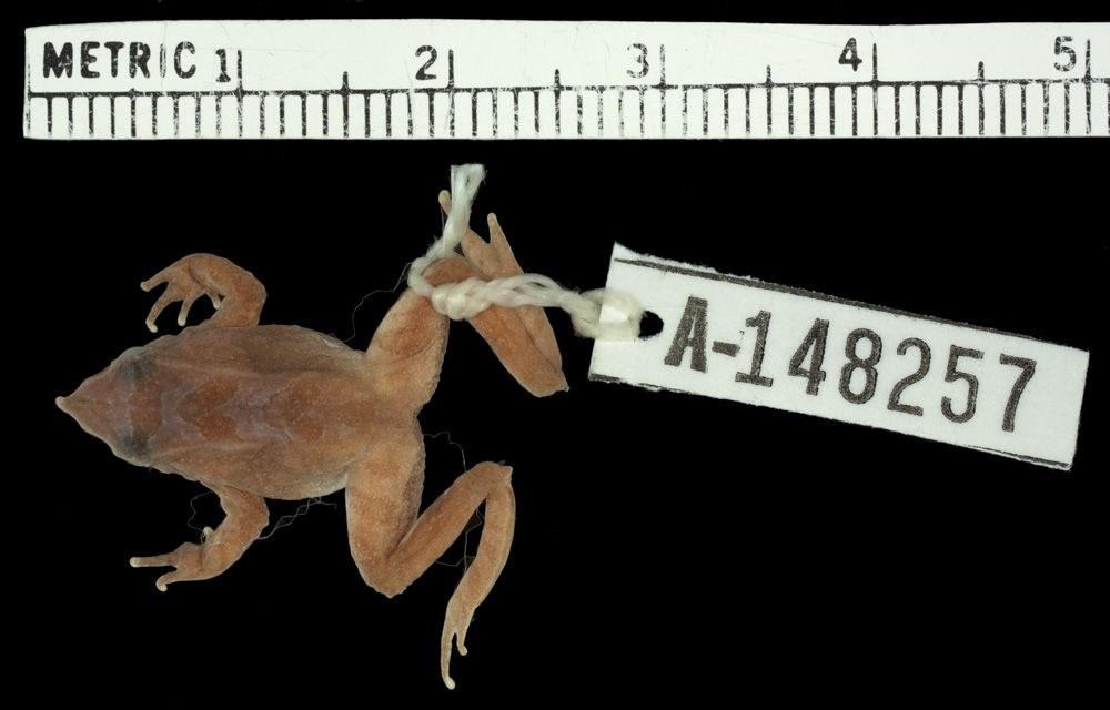
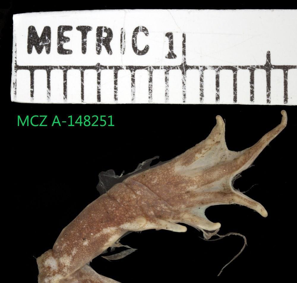
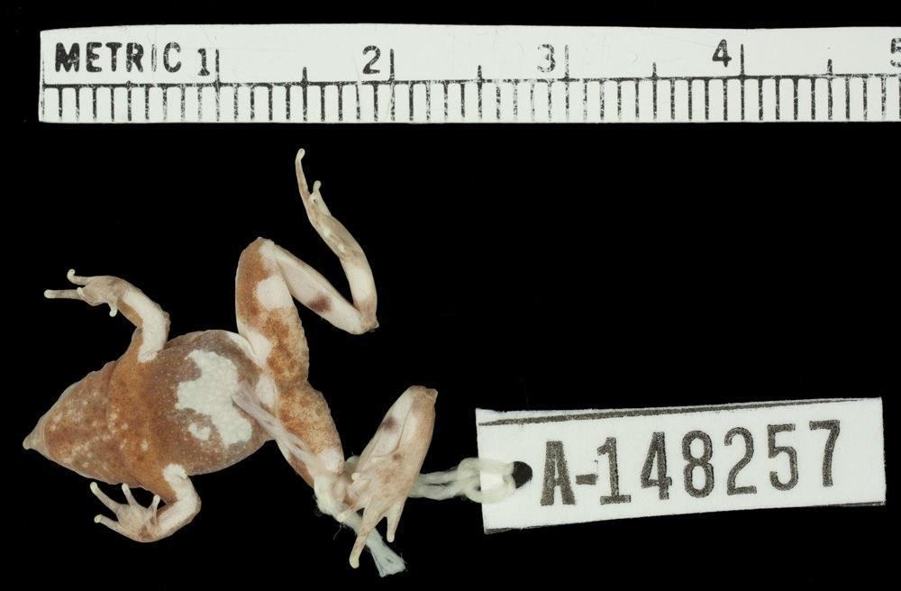
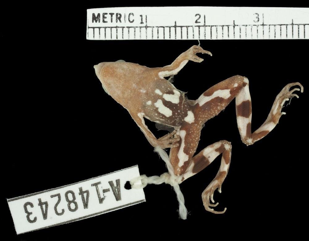
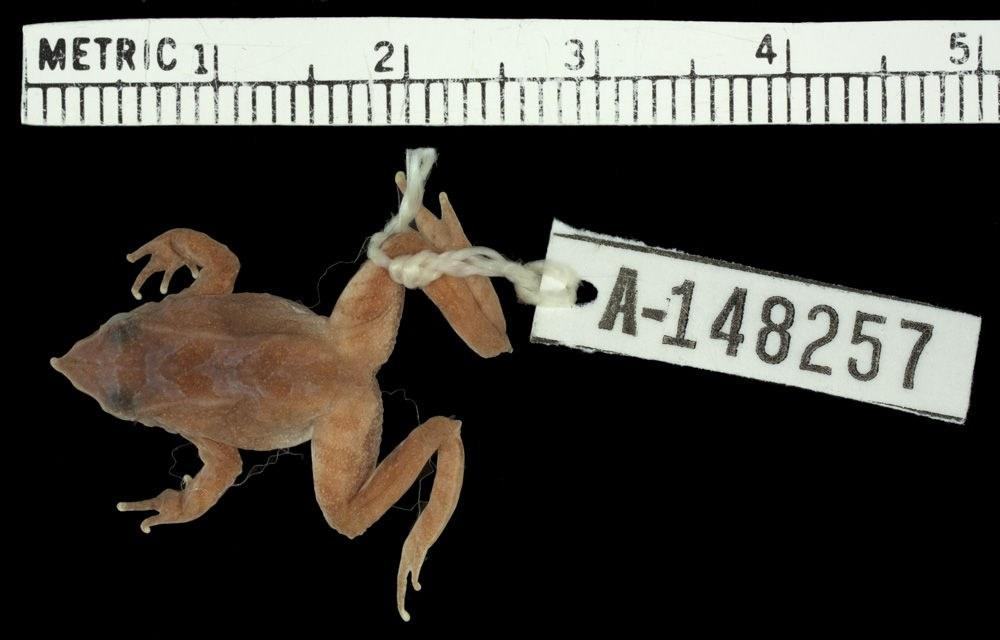
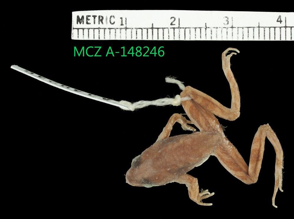
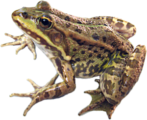
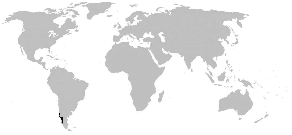
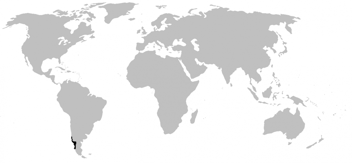
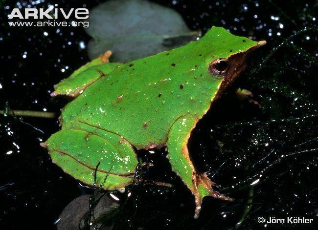
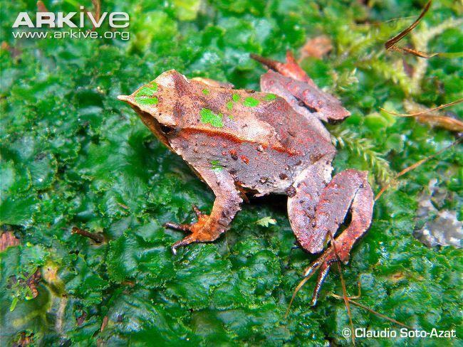
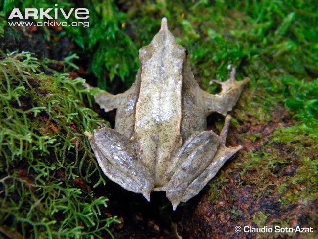
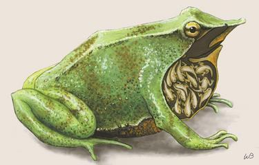
Rhinodermatidae – Mouth-brooding frogs Genus Rhinoderma Species Rhinoderma darwinii Species Rhinoderma rufum (Possibly extinct) The family is distributed along the southwestern coast of South America (Fig. 1). Members of the family are small in size (about 3 cm), live on land in moist habitats, have fleshy projections at the tip of the upper jaw (Fig. 2–4), webbing on the feet, and coloration in brown tones that may include green elements or be entirely green (Fig. 2–4). This family is characterized by a remarkable reproductive mechanism that sets them apart from other anurans. During reproduction, a small number of large eggs are laid by the female on waterlogged soil; in addition to the female, several males participate in guarding the clutch collectively until the tadpoles inside the eggs begin to wriggle and prepare to hatch. At that point, the males take up to fifteen eggs into their mouths and carry them in their vocal sacs (Fig. 5). When the tadpoles hatch, they lack external gills, have a poorly developed tail and jaws. Inside the vocal sacs they are nourished by the large yolk, and later attach to the inner wall of the vocal sac and feed at the expense of the male, as they lack an epidermal skin layer. By the time the tadpoles mature, the male transports them to temporary pools where they complete their development. Some researchers consider Rhinoderma rufum to be invalid and synonymize it with Rhinoderma darwinii. Rhinoderma rufum was last observed in the wild in 1980. It differs from Rhinoderma darwinii in having a less pronounced heel tubercle (Fig. 6–7), certain differences in coloration (Fig. 8–9), and a more prominent rostral projection (Fig. 10–11). Some systematists also include the family Rhinodermatidae within the family Cycloramphidae. Fig. 1: Range of Rhinodermatidae Fig.
2–4: Appearance and color variation of Rhinoderma darwinii Fig.
5: Tadpoles in the vocal sac of a male Fig.
6–7: Comparison of heel tubercles of Rhinoderma darwinii and Rhinoderma rufum Fig.
8–9: Comparison of ventral patterning in Rhinoderma darwinii and Rhinoderma rufum Fig.
10–11: Comparison of rostral projections of Rhinoderma darwinii and Rhinoderma rufum Sources: https://animaldiversity.ummz.umich.edu/ https://amphibiaweb.org/index.htm https://eol.org