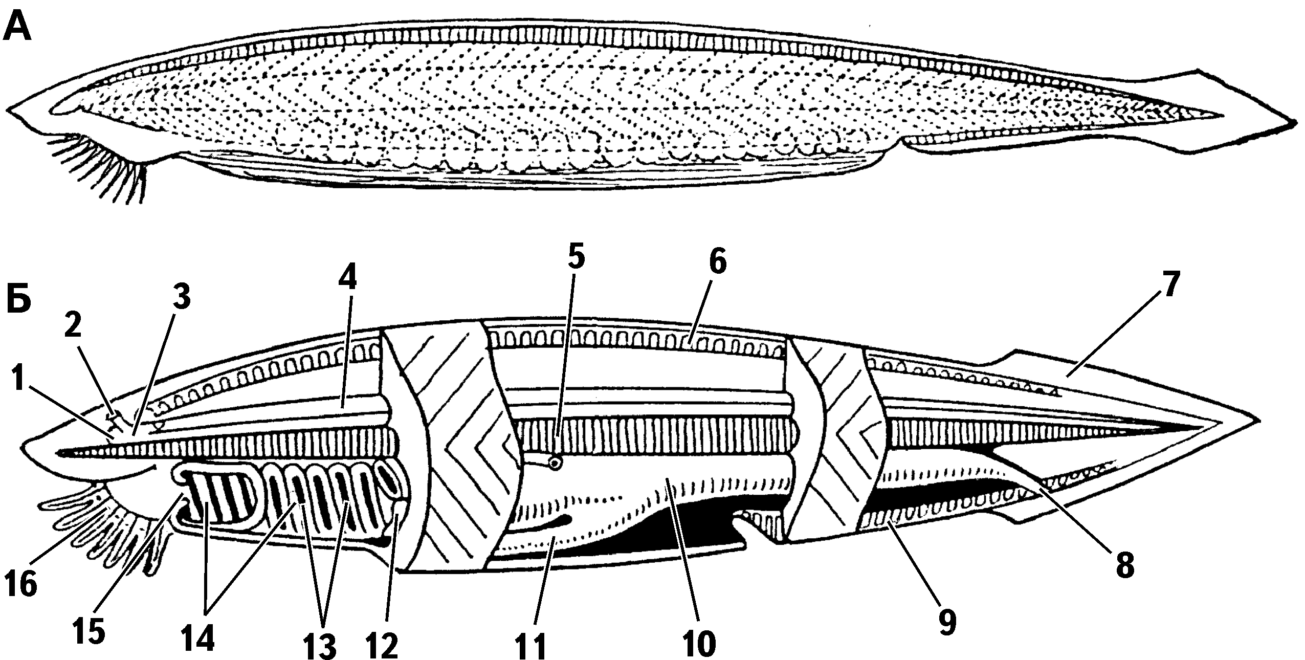
Structure of the lancelet: A – external appearance; B – structural diagram
1 – eye spot; 2 – olfactory pit; 3 – anterior end of the nerve tube; 4 – nerve tube; 5 – notochord; 6 – dorsal fin; 7 – caudal fin; 8 – anal opening; 9 – ventral fin; 10 – intestine; 11 – hepatic caecum; 12 – gonads; 13 – gill slits; 14 – pharynx; 15 – mouth; 16 – oral opening with tentacles
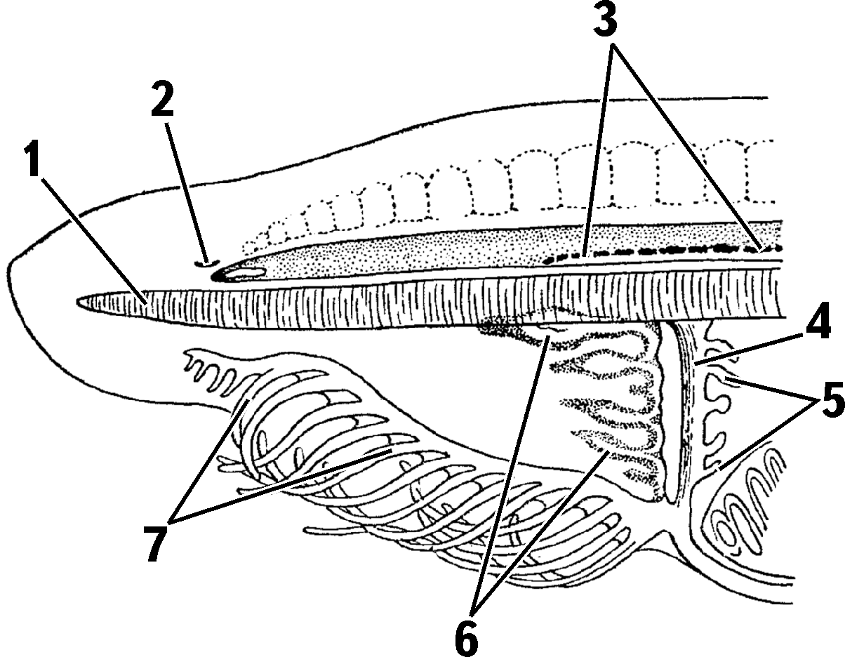
Head region of the lancelet
1 – notochord; 2 – olfactory pit; 3 – Hesse’s ocelli; 4 – velum; 5 – velar tentacles; 6 – ciliated organ; 7 – tactile tentacles
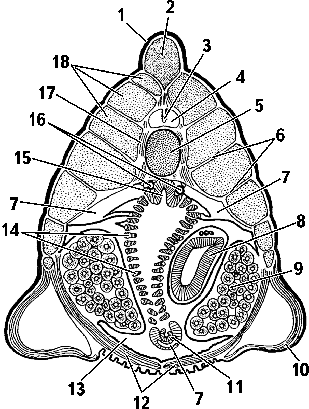
Cross section of the lancelet in the middle trunk region
1 – epidermis; 2 – dorsal fin; 3 – neurocoel; 4 – nerve tube; 5 – notochord; 6 – myosepta; 7 – coelomic canals; 8 – hepatic caecum; 9 – gonads; 10 – metapleural folds; 11 – endostyle; 12 – ventral muscle; 13 – atrial cavity; 14 – interbranchial septa; 15 – epibranchial groove; 16 – roots of the aorta; 17 – connective tissue tube; 18 – trunk musculature
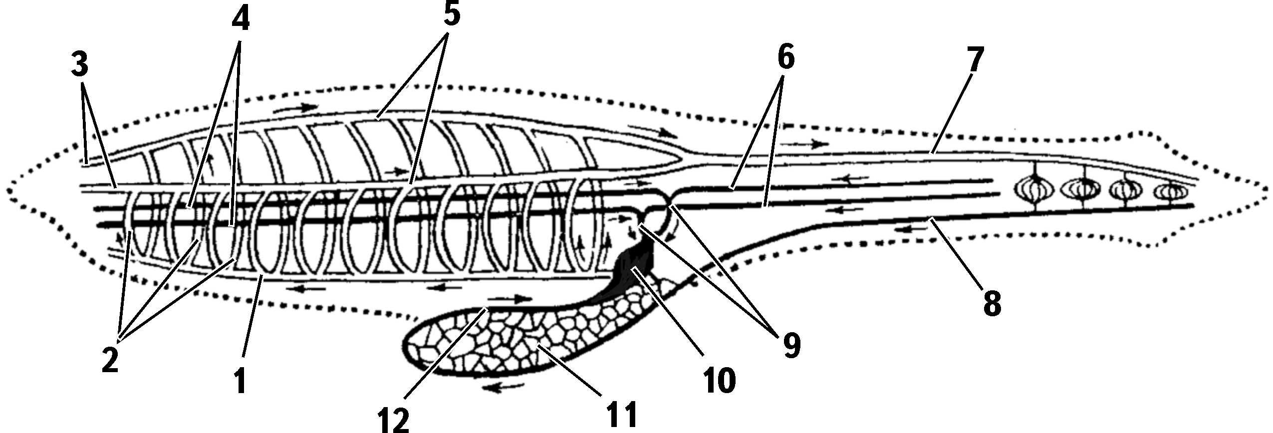
Circulatory system of the lancelet
1 – ventral aorta; 2 – branchial arteries; 3 – carotid arteries; 4 – anterior cardinal veins; 5 – roots of the aorta; 6 – posterior cardinal veins; 7 – dorsal aorta; 8 – subintestinal vein; 9 – Cuvierian ducts; 10 – venous sinus; 11 – portal system of the hepatic caecum; 12 – hepatic vein (arrows indicate direction of blood flow; veins are shown in black)
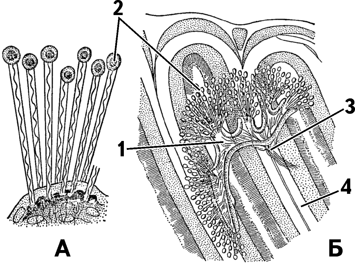
Nephridium (excretory tubule) of the lancelet: A – part of a nephridium with seven solenocytes; B – tubule with nephrostomes
1 – club-shaped solenocyte cells; 2 – openings into the body cavity (coelom); 3 – openings into the atrial cavity (with ciliated zone); 4 – gill slit
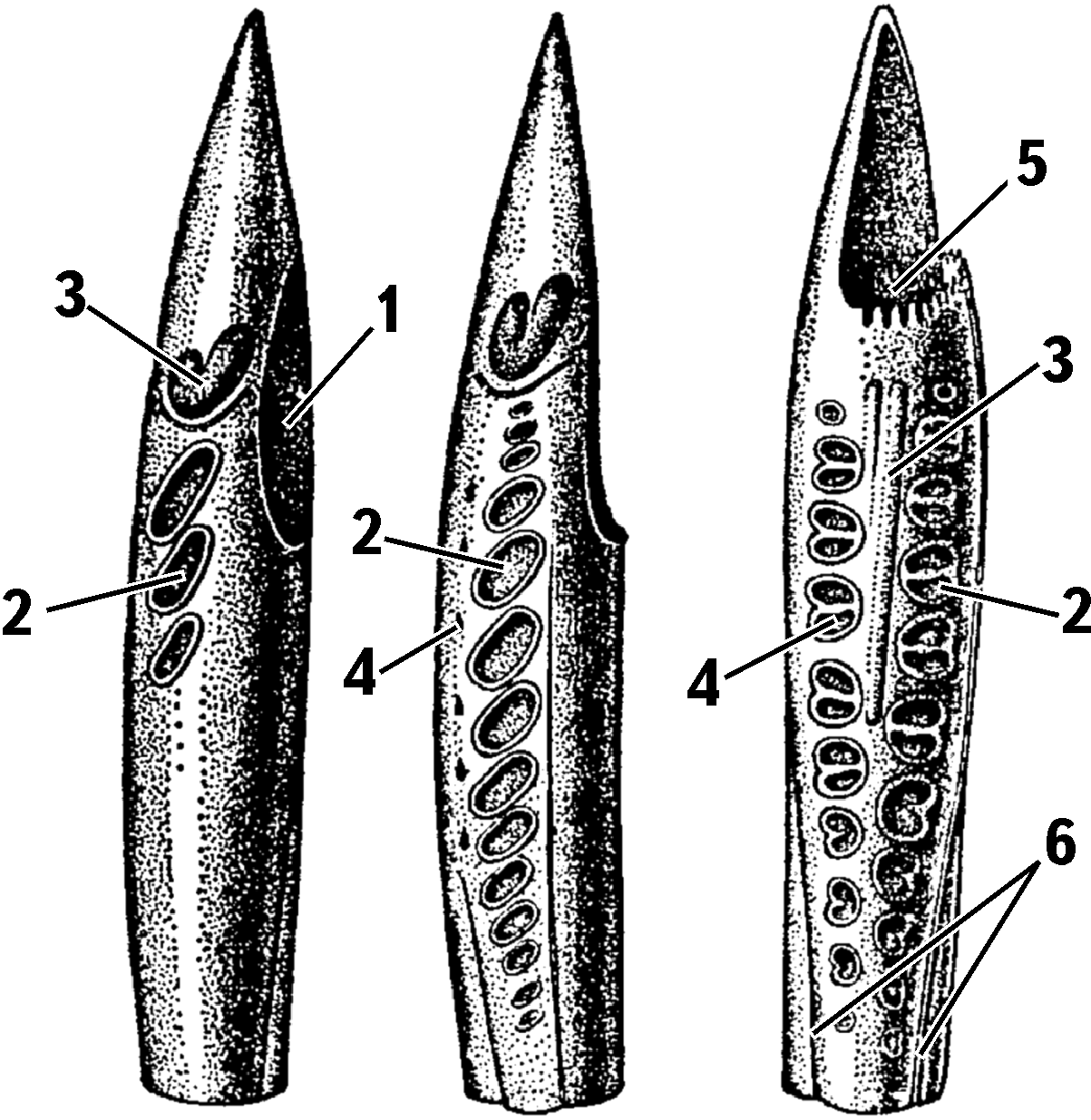
Successive development of gill slits in lancelet larva
1 – rudiment of the preoral pit; 2 – rudiment of gill slits on the left side; 3 – endostyle rudiment; 4 – rudiment of gill slits on the right side; 5 – rudiment of the mouth and larval mouth; 6 – metapleural folds, whose overgrowth forms the atrial cavity
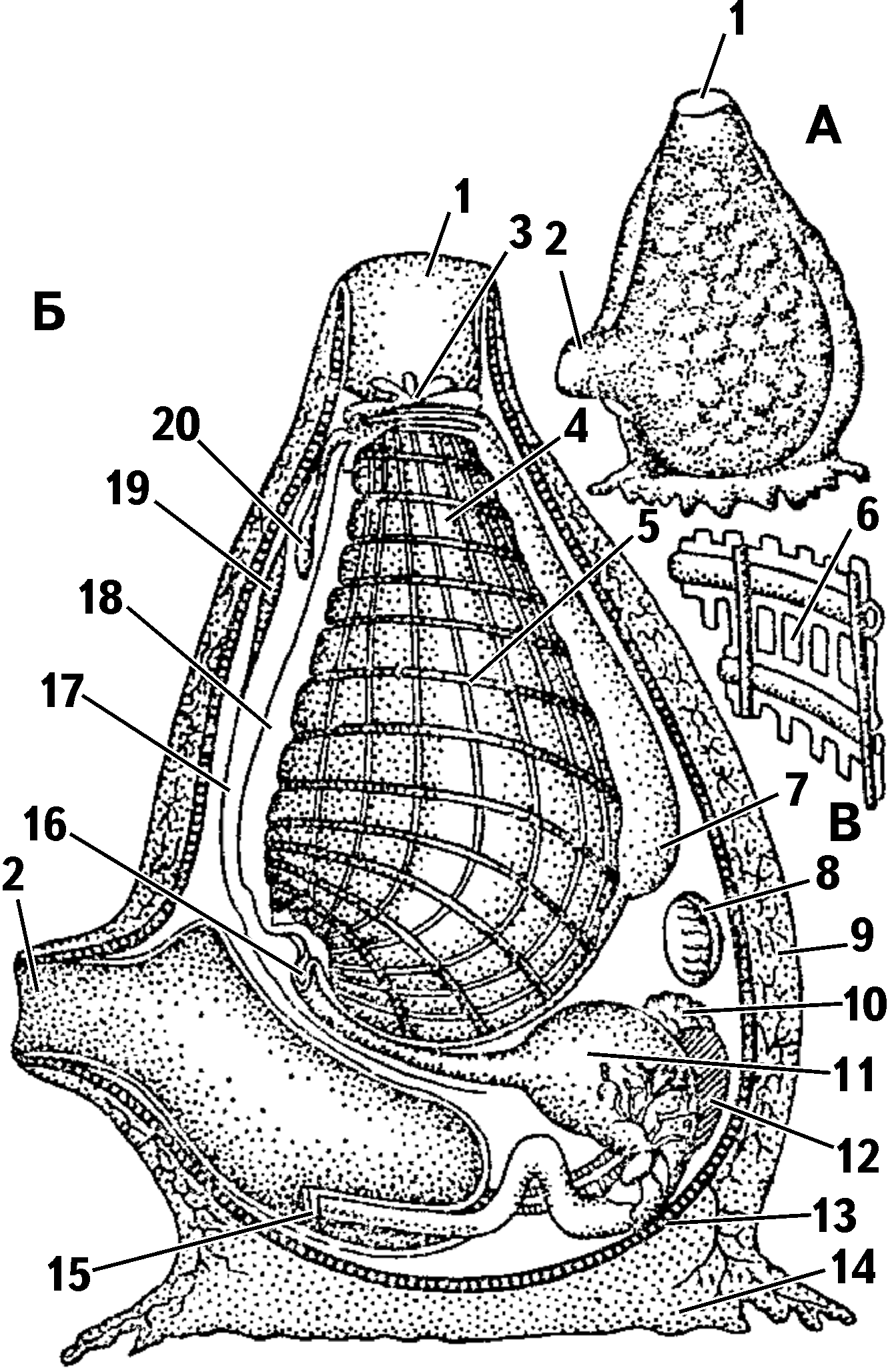
Structure of an ascidian: A – general appearance; B – longitudinal section; C – enlarged part of the pharyngeal wall with stigmata and vessels
1 – oral siphon; 2 – cloacal siphon; 3 – oral tentacles; 4 – pharynx; 5 – blood vessels; 6 – stigma; 7 – endostyle; 8 – heart; 9 – tunic; 10 – testes; 11 – stomach; 12 – ovaries; 13 – epithelium; 14 – base of ascidian; 15 – anal opening; 16 – beginning of esophagus; 17 – dorsal nerve trunk; 18 – dorsal groove; 19 – neural ganglion; 20 – subneural gland
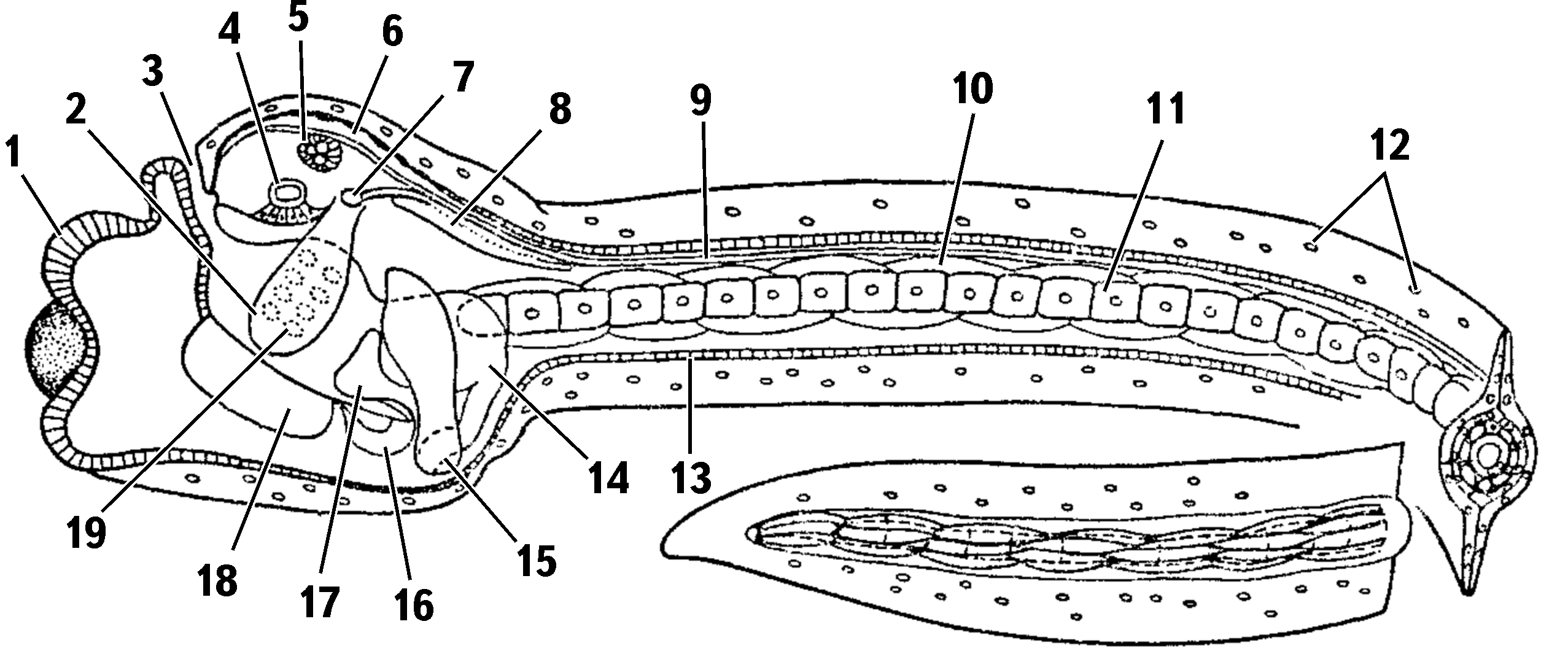
Diagram of ascidian larva structure
1 – sucker; 2 – rudiment of atrial cavity; 3 – mouth; 4 – statocyst; 5 – eye spot; 6 – brain vesicle; 7 – atriopore; 8 – visceral ganglion; 9 – nerve tube; 10 – muscle cells; 11 – notochord; 12 – free cells in the tunic; 13 – epidermis; 14 – stomach; 15 – intestine; 16 – heart; 17 – epicardium; 18 – endostyle; 19 – gill openings
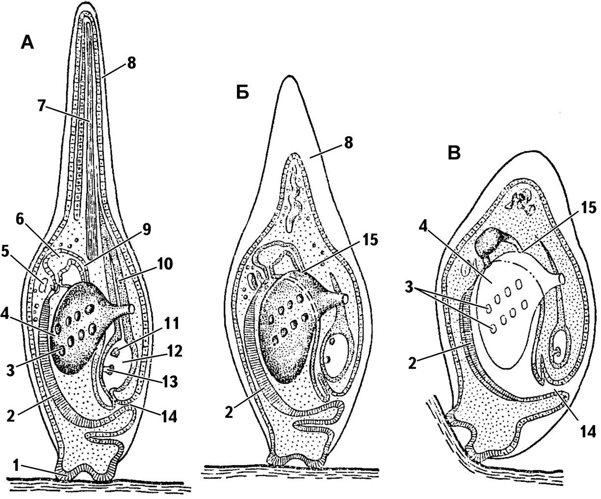
Successive stages of metamorphosis of the ascidian larva
1 – sucker; 2 – endostyle; 3 – gill openings; 4 – atrial cavity; 5 – heart; 6 – stomach; 7 – notochord; 8 – tail; 9 – intestine; 10 – nerve tube; 11 – eye spot; 12 – expansion of the nerve tube (brain vesicle); 13 – statocyst; 14 – mouth; 15 – anal opening
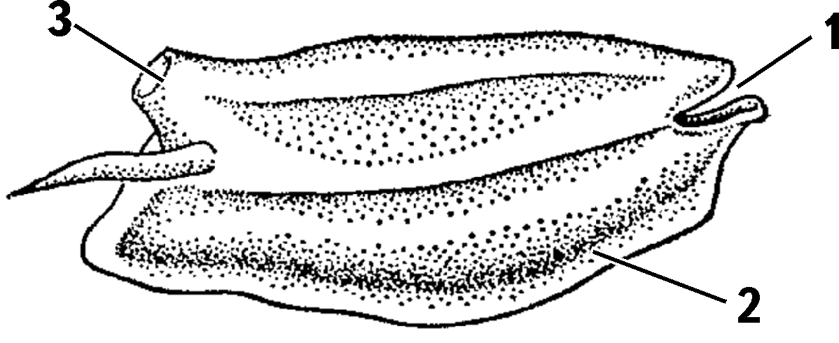
A solitary asexual individual of a salp
1 – mouth; 2 – ventral side; 3 – cloacal opening
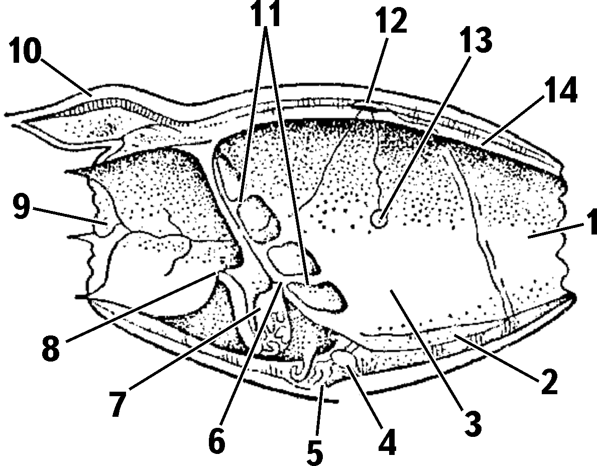
Longitudinal section of a doliolid
1 – oral siphon; 2 – endostyle; 3 – pharynx; 4 – heart; 5 – ventral stolon; 6 – esophageal opening; 7 – stomach; 8 – anus; 9 – cloacal siphon; 10 – dorsal stolon; 11 – septum with stigmata; 12 – neural ganglion; 13 – statocyst; 14 – muscle fibers
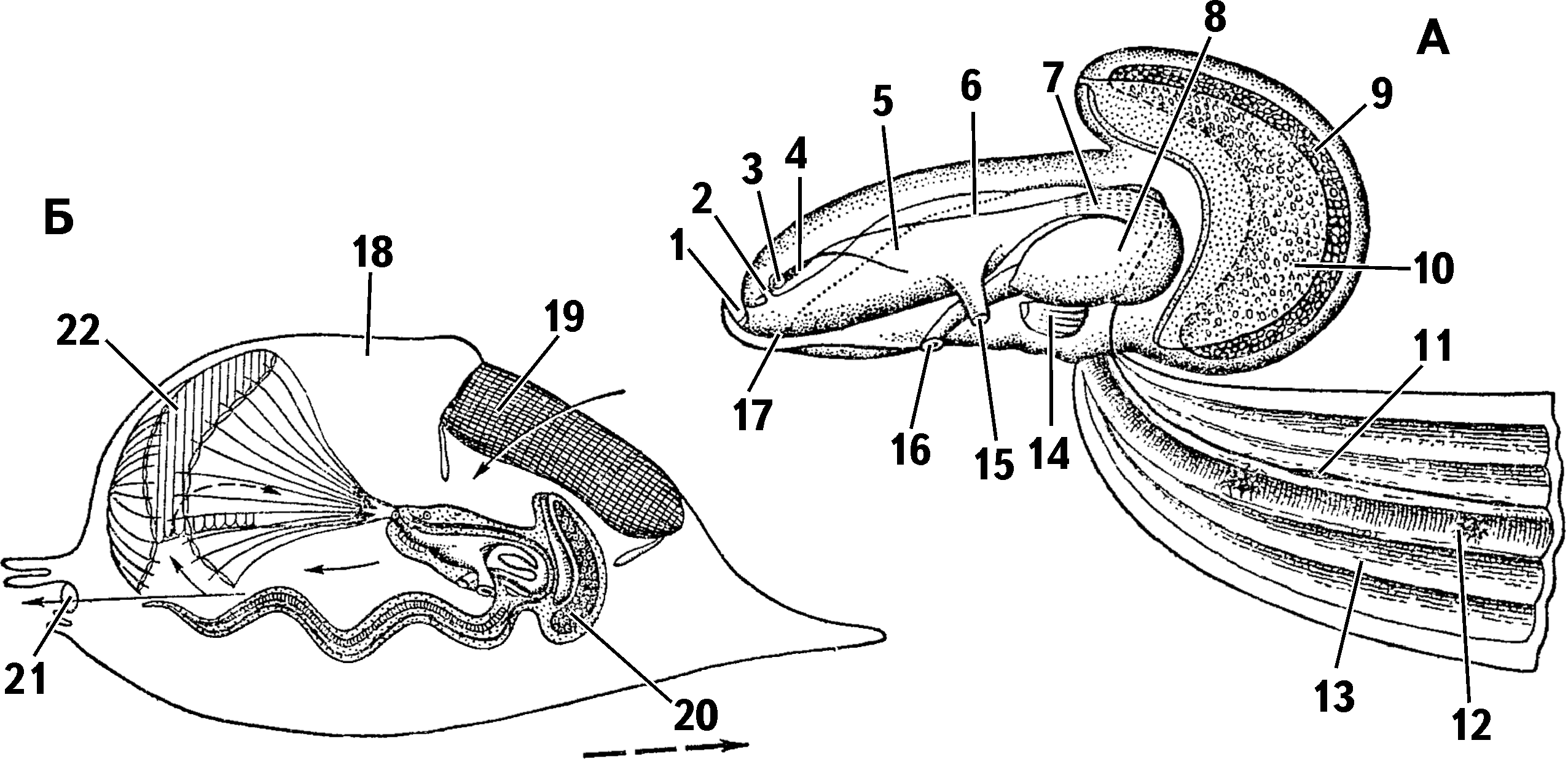
Appendicularian Oikopleura sp.: A – structure; B – animal in its house
1 – mouth; 2 – olfactory pit; 3 – statocyst; 4 – neural ganglion; 5 – pharynx; 6 – dorsal nerve trunk; 7 – esophagus; 8 – stomach; 9 – ovary; 10 – testis; 11 – thickening of the dorsal nerve trunk in the tail region; 12 – notochord; 13 – tail musculature; 14 – heart; 15 – gill opening – stigma; 16 – anus; 17 – endostyle; 18 – house; 19 – its screen; 20 – appendicularian; 21 – opening of the house; 22 – collecting net (thin arrows indicate the direction of water flow; dashed arrow – direction of movement of the house)