Usova, Shabanov (2009) On the optimization of the methodology for retrospective assessment of body size dynamics...
Justification of the methodology for studying the growth rates of green frogs, which our group currently uses. The age determination method has become more advanced since then, but it has not yet been described in detail. Usova E. E., Shabanov D. A. On the optimization of the methodology for retrospective assessment of size dynamics...
Usova E. E., Shabanov D. A. On the optimization of a method for retrospective assessment of body size dynamics of representatives of the Pelophylax esculentus complex (Amphibia, Ranidae) using skeletochronology // Zoocenosis-2009. Biodiversity and the role of animals in ecosystems. – Dnipro, DNU, 2009. – pp. 278–280.
UDC 597.851(477.54)
On the optimization of a method for retrospective assessment of body size dynamics of representatives of the Pelophylax esculentus complex (Amphibia, Ranidae) using skeletochronology
E. E. Usova, D. A. Shabanov
V.N. Karazin Kharkiv National University; e_usova@mail.ru; d.a.shabanov@gmail.com
Enhancing methods of the back‑calculated of the body size dynamics based on skeletochronology in specimens of Pelophylax esculentus complex (Amphibia, Ranidae).
O. E. Usova, D. A. Shabanov
V.N. Karazin Kharkiv National University; e_usova@mail.ru; d.a.shabanov@gmail.com
Retrospective skeletochronological assessment of growth of studied animal groups has not yet entered the widely accepted arsenal of methods in ecological and zoological research. From the authors’ point of view, even in the era of widespread molecular techniques, skeletochronological study of individual growth dynamics should be considered a highly promising method for investigating ontogenetic strategies of many vertebrate groups. The undervaluation of this method is due not only to the relatively (moderately) labor‑intensive preparation of histological sections required for retrospective growth assessment of individual specimens, but also to the lack of universally accepted and widely known protocols. Undeniable advantages of this method include the possibility of in‑vivo investigation, ease of obtaining and storing samples for research.
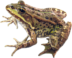
An example of a scientific problem for which skeletochronological data can be of unquestionable value is the study of the hybrid complex of Central European green frogs, Pelophylax esculentus complex. This complex consists of two parental species, the pool frog Pelophylax lessonae (Camerano, 1882) and the lake frog Pelophylax ridibundus (Pallas, 1771), as well as their hybrids, called edible frogs, Pelophylax esculentus (Linnaeus, 1758). The names of these forms follow the latest revision of amphibian taxonomy (Frost et al., 2006). Their more familiar traditional names are Rana lessonae Camerano, 1882, Rana ridibunda Pallas, 1771 and Rana esculenta Linnaeus, 1758. Hybrid frogs bear a separate name at the species level because they exhibit the phenomenon of hemiclonal inheritance, transmitting only one of the parental genomes to the gametes (Plötner, 2005 and other works). In several European regions, including Kharkiv Oblast, among hybrids diploids, triploids and even occasional tetraploids have been recorded (Borkin et al., 2004; Shabanov et al., 2006). Both diploid and triploid hybrids may differ in the type of gametogenesis: producing haploid or diploid gametes that carry one or the other genome. This leads to green frogs forming hemiclonal population systems (HPS): assemblages of co‑existing and interbreeding representatives of the parental species and various hybrid forms. In these HPS both clonal and ordinary (recombinant) genomes are transmitted. Interactions among different frog forms in HPS, as well as their varying viability, can cause changes in their composition over time (Shabanov et al., 2006). To elucidate the reasons for such dynamics, reliable data on lifespan, growth rates and age at sexual maturity of the different frog forms are needed. Skeletochronology is an adequate method for solving these tasks.
The authors performed skeletochronological study of growth rates in 49 representatives of the Pelophylax esculentus complex (Table 1).
Table 1. Studied material
(L – genome P. lessonae; R – genome P. ridibundus)
|
Form |
U | Use | Resource use |
juv. |
♀♀ |
♂♂ |
Total |
|
P. lessonae |
LL |
– |
1 |
– |
1 |
|
Aneuploid |
LLR |
– |
1 |
– |
1 |
|
P. esculentus |
LR |
– |
3 |
17 |
20 |
|
Aneuploid |
LRR |
10 |
3 |
3 |
16 |
|
P. ridibundus |
RR |
7 |
4 |
– |
11 |
|
Total |
17 |
12 |
16 |
49 |
|
In the examined specimens the fourth (longest) digit of the left hind limb was removed. The bone was cleared of soft tissue and decalcified. On a cryostat microtome transverse sections (20–22 µm thick) were obtained from the third phalanx (counting from the tip) at the mid‑diaphysis level, near the entry point of a blood vessel into the bone. Sections were photographed, and the smallest and largest transverse diameters of observable lines of sclerotic deposition (dark zones deposited in bone during hibernation) were measured. Considering that the first two lines of deposition undergo resorption and displacement by the endosteal cavity, the age of sexually mature frogs was determined by the number of fully observable lines of deposition plus 2.
E. M. Smirina (1983) determined changes in frog body length by constructing a regression line between body length and the area bounded by the corresponding line of deposition on the phalangeal section. This method was chosen based on the concept of an allometric relationship between body length and the diameter of tubular bones. Specialized studies (Marunouchi et al., 2000) showed that the most accurate estimation of frog body length is possible using a linear relationship between body length and bone diameter (Dahl‑Lea method). Similar relationships have been employed in recent works on lizards and frogs (Roitberg, Smirina, 2006; Zamaletdinov, Faizullin, 2008). We compared various methods of retrospective growth dynamics assessment on our material. Table 2 presents Pearson correlation coefficients and linear regression equations between frog body length (L) and the smallest (lastlmin) and largest (lastlmax) transverse diameters of the last line of deposition, the mean of these two values (lastlmed), and the area bounded by the last line of deposition (calculated using the ellipse area formula: lastS = lastlmin × lastlmax × π/4).
Table 2. Relationship of parameters of the last line of deposition on the phalangeal section to body length (parameter symbols as in text)
|
Parameter |
Correlation with L (µm) |
Linear regression equation |
|
lastlmin, µm |
r=0,9468 |
L=14,8+871,5×lastlmin |
|
lastlmax, µm |
r=0,9604 |
L=24,8+700,0×lastlmax |
|
lastlmed, µm |
r=0,9614 |
L=12,5+787,9×lastlmed |
|
lastS, µm2 |
r=0,9402 |
L=243,4+192,0×lastS |
As Table 2 shows, frog body length is most strongly correlated with the mean transverse diameter of the deposition line. The smallest transverse diameter is the most stable measurement against random deviation of the section plane from the bone axis. Nevertheless, this measure characterizes body length less accurately than the largest transverse diameter. Their averaged value (likely due to compensation of random variations of the two measures and measurement errors) proves to be the best parameter for further use. Importantly, the lines describing the relationship between body length and the linear parameters of the deposition line practically pass through the origin (i.e., correspond to a hypothetical frog with zero body length and zero finger phalanx thickness). The relationship between body length and the area of the deposition line has a different character.
Thus, for describing the studied frog assemblage one could use the relationship L = 788 × lastlmed. However, it should be taken into account that points representing individual specimens lie on the plane with axes lastlmed and L above or below this line (corresponding to “stocky” or “slender” frogs). The position of frogs on this plane reflects developmental peculiarities of the studied forms as well as sexual and individual traits. Logically, it can be assumed that frogs situated above or below the overall regression line follow a line originating from the origin that is above or below the overall line. Based on this assumption, one can conclude that L/ lmed = iL/ ilmed, where lmed is the averaged transverse diameter of the last deposition line (or the finger phalanx as a whole) at the time of study of a frog of length L, iL is the body length at the i‑th year of life, and ilmed is the averaged transverse diameter of the i‑th deposition line. From this, we can establish the final formula for retrospective determination of body length of the studied green frog specimens: iL = L × ilmed / lmed. This method aligns with approaches used in other studies (Roitberg, Smirina, 2006; Zamaletdinov, Faizullin, 2008), is easy to interpret, is based on a parameter closely linked to body length, and allows accounting for individual variation of each specimen.
The work was supported by a joint grant of the Ukrainian Science Foundation and the Russian Foundation for Basic Research.