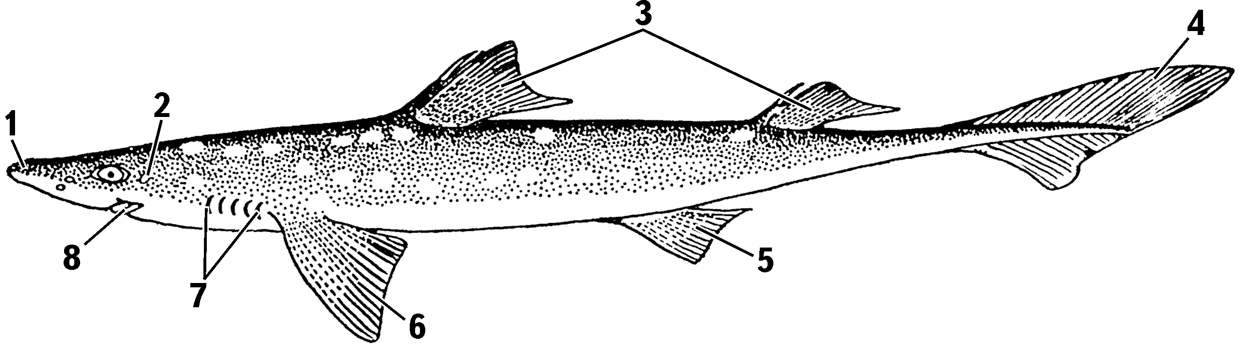
Shark body, lateral view
1 — rostrum; 2 — spiracle; 3 — dorsal fins; 4 — caudal fin; 5 — pelvic fin; 6 — pectoral fin; 7 — gill-slit openings; 8 — mouth opening
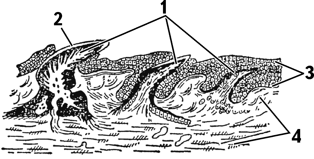
Longitudinal section through shark skin and placoid scale
1 — placoid scales at different stages (black — dentine, white — pulp); 2 — enamel layer; 3 — epidermis; 4 — corium
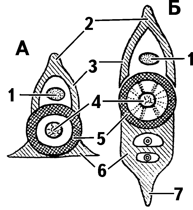
Structure of shark vertebrae: A — trunk vertebra; B — caudal vertebra
1 — spinal canal; 2 — upper spinous process; 3 — upper arches; 4 — notochord remnant; 5 — vertebral body; 6 — lower arches; 7 — lower spinous process
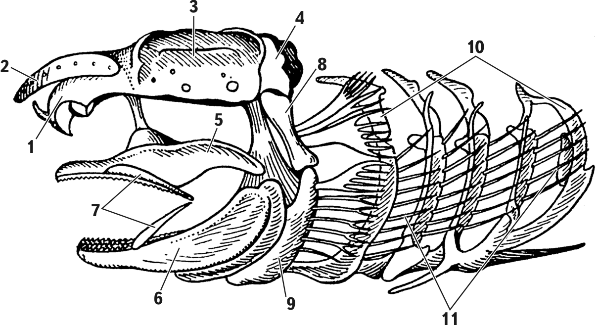
Shark skull, lateral view
1 — olfactory capsule; 2 — rostrum; 3 — orbit; 4 — auditory capsule; 5 — palatoquadrate cartilage; 6 — Meckel’s cartilage; 7 — labial cartilages; 8 — hyomandibula (suspensorium); 9 — hyoid; 10 — gill arches; 11 — rays supporting interbranchial septa

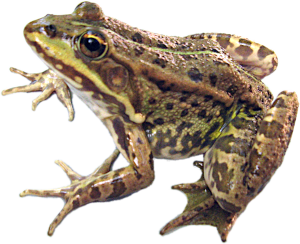
Relations of anterior visceral arches to the axial skull: A — amphistylic shark skull (Heptanchus); B — hyostylic shark skull; C — amphistylic skull of a bony ganoid (Amia); D — autostylic skull of a terrestrial vertebrate (frog)
1 — hyomandibula; 2 — hyoid; 3 — ligament; 4 — Meckel’s cartilage; 5 — lower jaw; 6 — otic process; 7 — ligament; 8 — palatoquadrate cartilage
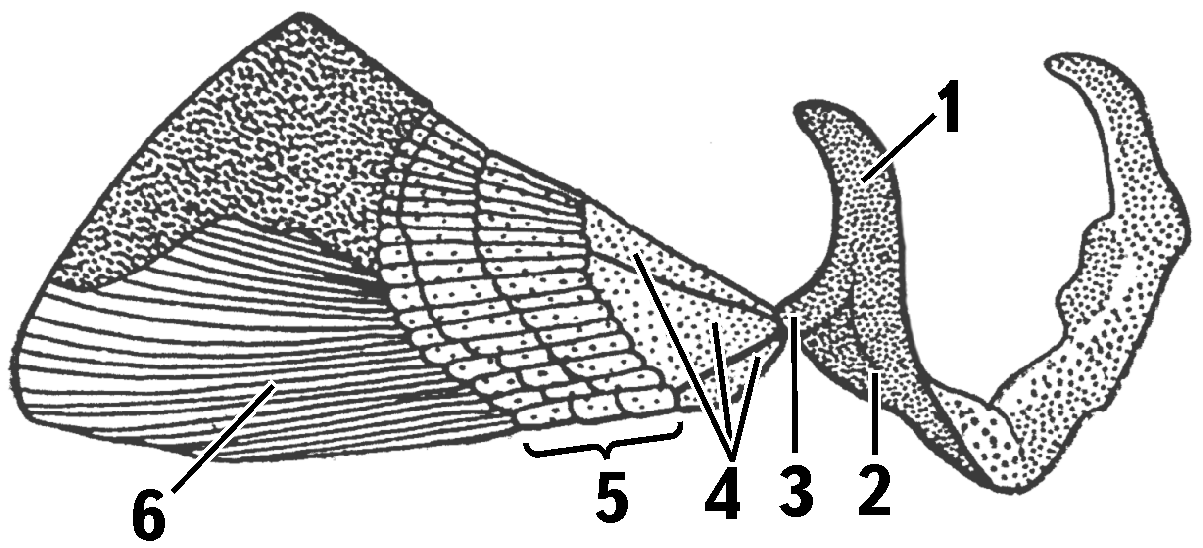
Pectoral girdle and pectoral fin of a shark
1 — scapular part; 2 — coracoid part; 3 — articular process; 4 — basal fin cartilages; 5 — radial cartilages; 6 — elastoidin fibers
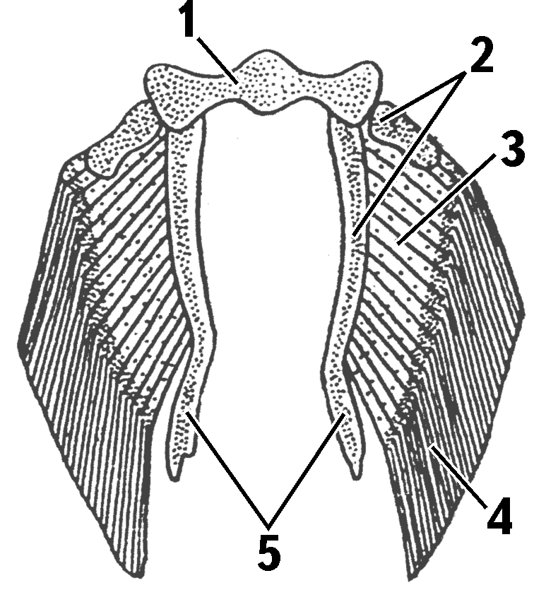
Pelvic girdle and pelvic fins of a male shark
1 — pelvic girdle; 2 — basalia; 3 — radials; 4 — elastoidin fibers; 5 — skeleton of copulatory organ
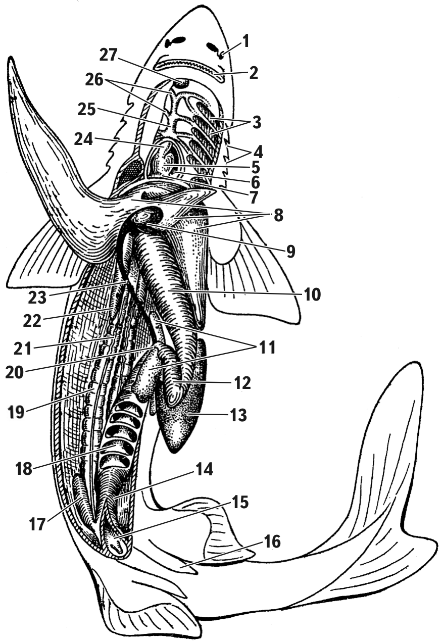
Dissected shark (male)
1 — nostril; 2 — mouth cleft; 3 — gills; 4 — external gill-slit openings; 5 — ventricle; 6 — atrium; 7 — sinus venosus; 8 — liver; 9 — gall bladder; 10 — cardiac stomach; 11 — pancreas; 12 — pyloric stomach; 13 — spleen; 14 — rectum; 15 — cloaca; 16 — pelvic-fin claspers; 17 — rectal gland; 18 — opened large intestine with spiral valve; 19 — kidney; 20 — small intestine; 21 — vas deferens; 22 — testis; 23 — bile duct; 24 — conus arteriosus; 25 — ventral aorta; 26 — afferent branchial arteries; 27 — thyroid gland
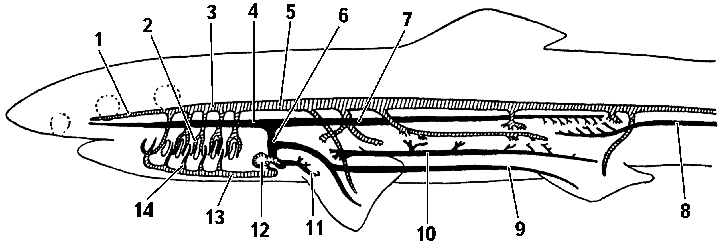
Shark circulatory system (left vessels shown from paired vessels)
1 — carotid artery; 2 — efferent branchial vessels; 3 — left aortic root; 4 — left anterior cardinal vein; 5 — dorsal aorta; 6 — left Cuvierian duct; 7 — left posterior cardinal vein; 8 — caudal vein; 9 — left lateral vein; 10 — hepatic portal vein; 11 — hepatic vein; 12 — heart; 13 — ventral aorta; 14 — afferent branchial artery (veins in black, arteries hatched)
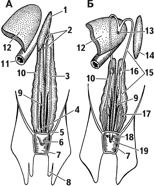
Urogenital system of cartilaginous fishes: A — male; B — female
1 — left testis (right not shown); 2 — efferent ducts; 3 — vas deferens; 4 — seminal vesicle; 5 — sperm sac; 6 — urogenital papilla; 7 — cloacal cavity; 8 — pelvic-fin clasper; 9 — ureter; 10 — kidney; 11 — esophagus; 12 — liver; 13 — common funnel of both oviducts; 14 — left ovary (right not shown); 15 — oviduct; 16 — shell gland; 17 — uterus; 18 — oviduct opening; 19 — urinary papilla
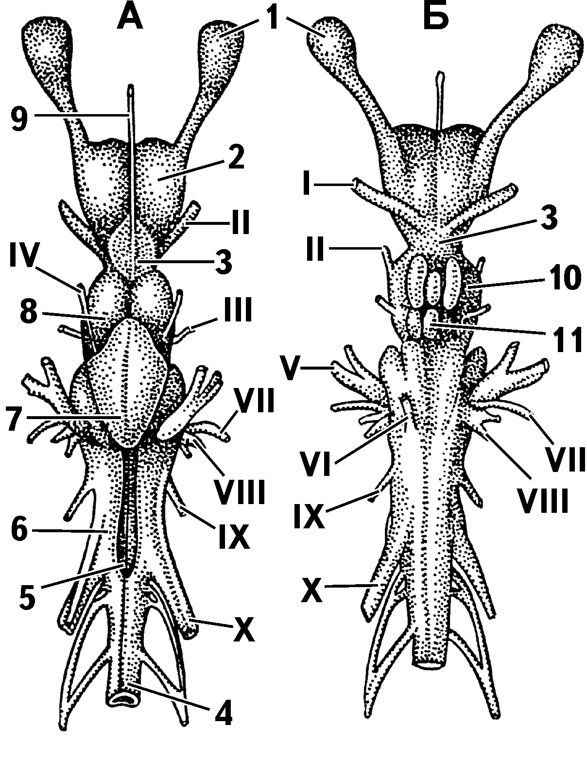
Spiny dogfish brain: A — dorsal view; B — ventral view
1 — olfactory bulb; 2 — forebrain; 3 — diencephalon; 4 — spinal cord; 5 — cavity of the fourth ventricle; 6 — medulla oblongata; 7 — cerebellum; 8 — optic lobes of the midbrain; 9 — pineal organ; 10 — midbrain; 11 — pituitary gland (Roman numerals indicate cranial nerves)
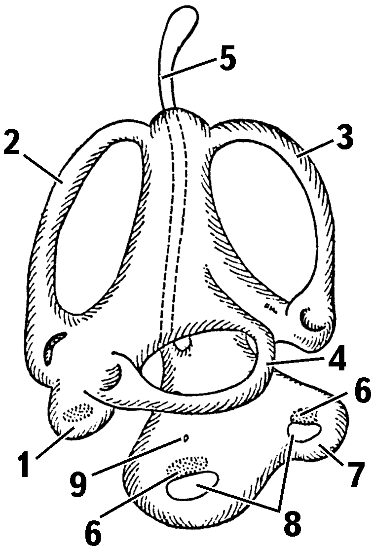
Scheme of fish inner ear
1 — utriculus; 2–4 — semicircular canals; 5 — endolymphatic duct; 6 — sensory fields; 7 — lagena (hollow protrusion of the sacculus); 8 — otoliths; 9 — sacculus
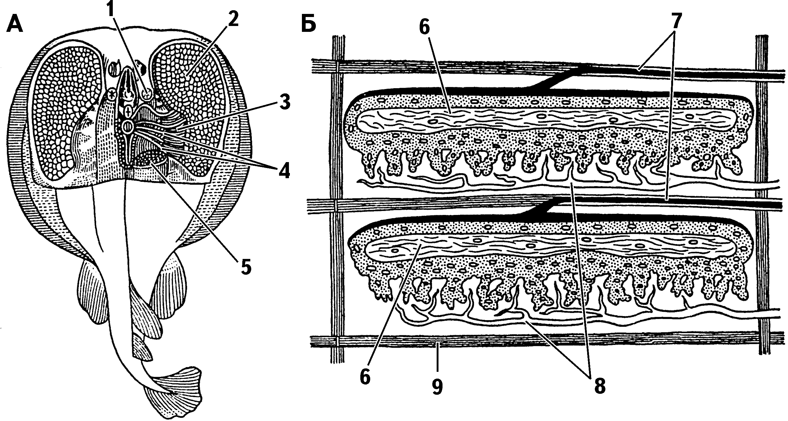
Electric ray — Torpedo marmorata: A — dissected electric organs and brain; B — structure of electric organ: two electric plates
1 — spiracle; 2 — electric organ; 3 — gill slits; 4 — main nerves to electric organ; 5 — cut muscle; 6 — central layer; 7 — blood vessels; 8 — nerves approaching organ; 9 — gelatinous connective-tissue sheath